Theme 3 - Case Studies
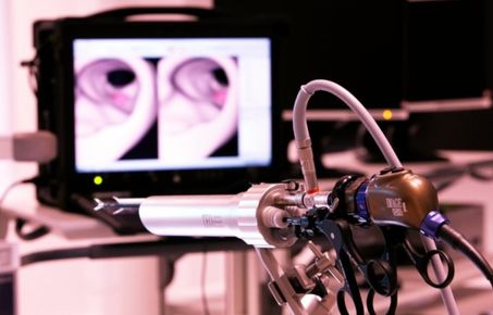
High-speed line-scan confocal laser endomicroscope (LS-CLE)
Probe based confocal laser endomicroscopy (p-CLE) is a novel optical biopsy tool that allows real time in vivo histological imaging of tissue surface without having to section or fix it. The currently widely used Cellvizio p-CLE system offers tissue imaging with microscopic resolution and has demonstrated promising sensitivity and specificity in small clinical studies primarily conducted by academics in research settings.
A striking advantage of LS-CLE systems over confocal endomicroscopes like Cellvizio and other microscopy platforms, is its ability to acquire confocal images at high acquisition rates. This allows examination of large anatomical areas like the entire surface of surgically excised specimens and its margin status in a timely manner with microscopic resolution.
.jpg) However, progression of p-CLE as an intra-operative tool for surgical applications has not reached its maximum potential. They have been limited in their capabilities of imaging in vivo tissue for the following reasons:
However, progression of p-CLE as an intra-operative tool for surgical applications has not reached its maximum potential. They have been limited in their capabilities of imaging in vivo tissue for the following reasons:
- slow image acquisition rate 10-12 fps,
- small field of view, 0.25 to 0.8 mm, depending on the selection of the distal optics, and
- restriction to use single fluorescent agent due to due to low-speed, single excitation, and single fluorescence spectral band imaging system.
The slow speed and small field of view makes it difficult for surgeons to maintain adequate probe-tissue contact and dexterity while scanning inside large-areas like breast cavities, especially during manual scanning.
Our research team recently developed a high-speed line-scanning confocal endomicroscope (LS-CLE) that can achieve frame rates up to 120 fps, an order of magnitude improvement over existing commercially available p-CLE systems like Cellvizio. The optical system has been changed slightly to increase the image acquisition rate.
Instead of implementing a point-by-point scanning mechanism to capture a 2D image across the tissue, a line is scanned in one direction and captured by a high-speed rolling-shutter CMOS camera. The rolling shutter of the CMOS camera operates as a virtual detector slit that rejects most of the out-of-focus light leading to optical sectioning at frame rates of 120 Hz.
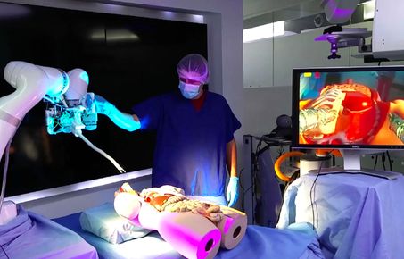
The high-speed LS-CLE system we developed could be easily adapted to suit different applications. Two particular clinical areas are bladder and breast cancer.
Bladder
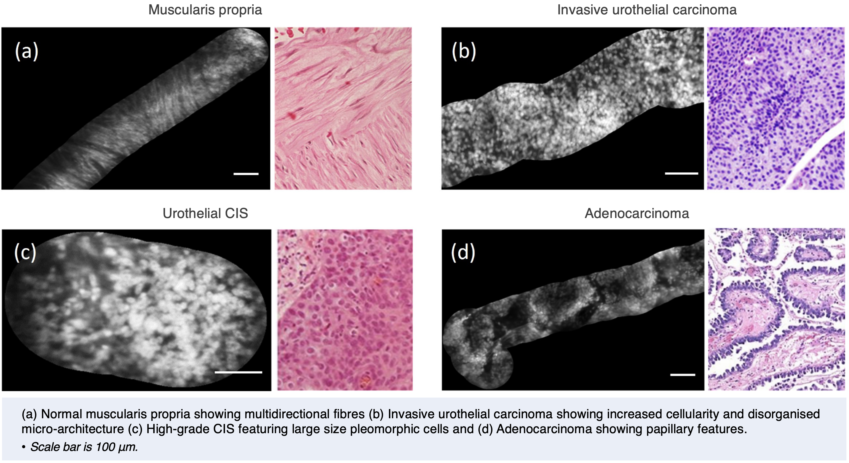
- Urothelial carcinoma is a common malignancy of the urinary tract, with the majority of these tumours being located in the bladder. Bladder cancer patients with superficial tumours are associated with a significantly higher risk of developing local recurrence and disease progression (70%), necessitating regular endoscopic surveillance, as well as a risk of superficial disease (30%) progressing into the more significant muscle-invasive state.
- Existing intra-operative techniques based on white light cystoscopy (WLC) lacks the ability to provide histopathologic information, which is essential for early detection, diagnosis and prognosis, while other developments such as blue light photodynamic diagnosis have lacked the specificity/sensitivity for widespread clinical adoption.
Breast cancer
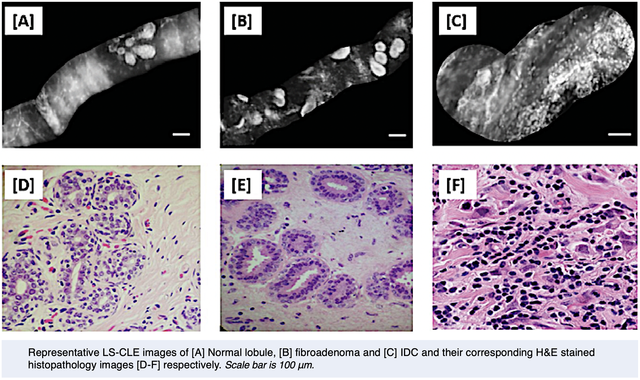
- Breast cancer is one of the leading causes of cancer in women, accounting for 23% of the total cancer cases and 14% of all cancer-related female deaths. It is also the most common cancer in the United Kingdom (UK) with more than 55,000 new diagnoses each year. Surgery to remove the tumour is part of the primary treatment in 81% of breast cancer patients.
- Breast conserving surgery (BCS) targets the excision of the tumour with a surrounding margin of normal tissue. Whilst BCS in combination with adjuvant radiotherapy has been demonstrated to have a similar overall survival rate to mastectomy, there are still concerns that conservative resection may not provide complete tumour excision.
- A key factor for future recurrence of breast cancer is the status of the microscopic margins of the resected breast specimen. Despite improvement in localisation techniques, up to 20% of patients will have positive or close margins on histology, mandating a second operation of re-excise further tissue along with many negative consequences like increased risk of postoperative infections, poor cosmesis, patient trauma and increase medical costs due to a longer stay in hospital.
- Intra-operatively, gross examination by palpation of specimen by the surgeon is not a reliable method of assessing clear surgical margins as evidenced by positive tumour margins of 50% if used alone. Most often these specimens are analysed post-operatively, resulting in at least 48 hours delay from the time of surgery to the provision of histopathology results.
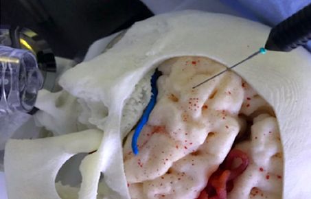
Our LS-CLE system allows for non-invasive real-time ‘virtual’ histology imaging of whole freshly excised breast and bladder tissue specimens without having to section and fix them. Our preliminary studies demonstrate the potential of using LS-CLE as a flexible endomicroscope in identifying discernible features corresponding to normal, benign and neoplastic bladder urothelium and breast tissue.
Case for Bladder
- LS-CLE mosaics and H&E stained histology of various pathologies of urinary tract
Case for Breast Cancer
- Rapid Digital Histology of Early Breast Cancer
(Histopathological analysis of excised tissue during oncological resection)
It is the first step towards recognising morphological appearances specific to micro-confocal imaging of these tissue specimens and may prove to be a valuable addition to routine surgical imaging when used in future translational studies such as delineating specific types of urothelial carcinomas, assessing the margin status of whole wide local excision specimens, scanning the breast cavity as well integration of LS-CLE with robotically assisted procedures.
- Khushi Vyas, Eric Yeatman, and Ranan Dasgupta, "Rapid digital histology of urothelial carcinoma using line-scan confocal laser endomicroscopy", Biophotonics Congress: Biomedical Optics 2022 (Translational, Microscopy, OCT, OTS, BRAIN), April 2022.
- Khushi Vyas, Ahmed Ezzat, Martin Asenov, Manish Chauhan, Subramanian Ramamoorthy, Animesh Jha, Daniel Leff, "Line-scan Confocal Endomicroscopy for Rapid Digital Histology of Early Breast Cancer", in Conference on Lasers and Electro-Optics, Technical Digest Series (Optica Publishing Group, 2022), paper ATh4I.7, May 2022.
- Ahmed Ezzat, Khushi Vyas, Martin Asenov, Manish Chauhan, Animesh Jha, Subramanian Ramamoorthy, Daniel Leff, "Portable confocal endomicroscopy for ductal feature characterization: Toward margin assessment in breast-conserving surgery", The American Society of Breast Surgeons Official Proceedings, Volume XXIII 2022 Annual Meeting Scientific Session Abstracts, Ann Surg Oncol 29 (Suppl 1), 190 (1148279), April 2022.
Theme 3 Highlights
Research Outcomes
- To know more about our Research Outcomes (Key Findings, Research Databases and Models, Research Tools and Methods, Collaboration, Software and Technical Products, Intellectual Property and Engagement Activities), please visit UKRI - Micro-Robotics for Surgery (EP/P012779/1) website.